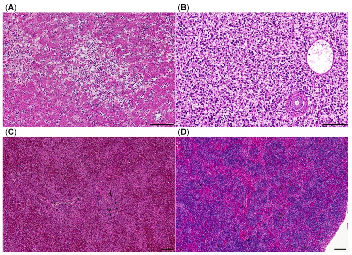
Figure 4.
Histology of brown trout liver and spleen, HE stain, (A) liver with multifocal liquefactive necrosis (center) and viable hepatocytes without vacuolation suggesting depletion of glycogen stores; (B) liver from a healthy brown trout; (C) congested spleen with lymphocytic depletion of white pulp areas; (D) spleen from a healthy brown trout; bars = 100 µm.

