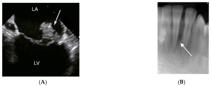
Figure 3.
Imaging signs of infections and inflammation that may be associated with A. actinomycetemcomitans. (A) Transesophageal echocardiography of a heart with mitral valve infective endocarditis. The arrow marks a large vegetation on the posterior leaflet between left atrium (LA) and left ventricle (LV); usually, vegetations caused by A. actinomycetemcomitans are of smaller size. (B) 14-year old girl of African ethnicity. The radiograph shows an extensive and apparently rapid loss of the periodontal support of the lower incisor 31. Pictures by courtesy of close clinical collaborators of the authors.

