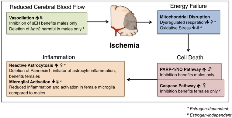
Figure 2.
Schematic representation of sex differences in stroke pathology and therapeutic efficacy. Sex differences in mitochondrial disruption exist; experimental stroke models in both mice and rats have shown that female animals experience less oxidative stress and dysregulated respiration after ischemia than males, an advantage that is eliminated by ovariectomy or aging. Sex differences in vasodilation have also been described, in which experimental mouse models have shown that females experience enhanced vasodilation at baseline compared with males, and male animals show benefit only when vasodilation is enhanced [inhibition of soluble epoxide hydrolase (sEH), selection of angiotensin II type 2 receptor (Agtr2)]. Studies in mice have shown enhanced reactive astrocytosis in females after ischemic, which can be ameliorated by the deletion of the proinflammatory protein Pannexin 1. Conversely, female mice show reduced microglial activation compared with male animals in experimental mouse models of ischemic stroke. In vivo and in vitro studies in mice and mouse-derived neuronal cells have demonstrated that the PARP-1/nitric oxide (NO) pathway of cell death predominates in males, whereas the caspase pathway of cell death is dominant in females. In line with this, inhibition of each of these pathways benefits only males, or only females, respectively.

