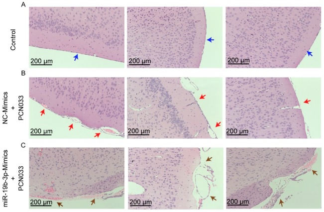
Figure 6.
Histopathological analysis of meninges in PCN033-challenged mice with or without miR-19b-3p mimic pretreatment. (A) The normal meninges from uninfected mice (blue arrows). (B) The meninges from PCN033-challenged mice receiving the control mimics. Red arrows indicate meninges thickening, hyperemia, and inflammatory cell accumulation. (C) The meninges from PCN033-challenged mice receiving miR-19b-3p mimic pretreatment, and brown arrows show more severe thickening, hyperemia, and accumulation of inflammatory cells, as compared to the challenged brains with control mimic treatment. Scale bar = 200 μm.

