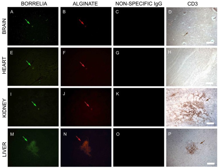
Figure 9.
Representative IHC images of Borrelia, alginate, and CD3+ T lymphocytes staining in infected brain, heart, kidney, and liver autopsy tissue sections. Tissues sections that had positive staining for Borrelia (green staining: A,E,I,M) and alginate (red staining: B,F,J,N) were subjected to additional IHC analyses by immunostaining the sequential sections with a T cell marker, CD3-specific antibody (brown staining: D,H,L,P). Fluorescent images were taken at 100x magnification to illustrate a larger section of the tissue. CD3-positive lymphocytes surrounded these aggregates, as depicted with brown staining in the brain, kidney, and liver tissues (D,L,P). There was no presence of CD3-positive lymphocytes in the heart tissues (H). Nonspecific IgG antibody was used as a negative control for the primary antibodies (C,G,K,O). Scale bar: 100 μm.

