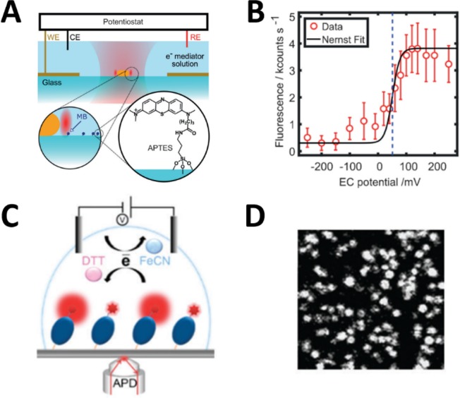
Figure 2.

Detection of surface-immobilized single molecules on an electrode. (A) Schematic illustration of the combined electrochemical–confocal setup with immobilized AuNRs (not to scale) and MB molecules on the modified glass surface. (B) Ensemble fluorescence response of around 260 unenhanced MB molecules versus electrochemical potential. The black curve is Nernst fit, and the dashed line indicates the obtained midpoint potential. (A), (B) Reproduced from Zhang, W.; Caldarola, M.; Pradhan, B.; Orrit, M. Angew. Chem. Int. Ed. 2017, 56, 3566–3569 (ref (17)). Copyright 2017 Wiley-VCH. (C) Schematic illustration of the detection of single azurin-Cy5 molecules immobilized on a modified glass surface using confocal microscopy. (D) A confocal fluorescence image of immobilized azurin-Cy5 molecules on glass. (C), (D) Reproduced from Akkilic, N.; Van Der Grient, F.; Kamran, M.; Sanghamitra, N. J. M. Chem. Commun. 2014, 50, 14523–14526 (ref (16)). Copyright 2014 Royal Society of Chemistry.
