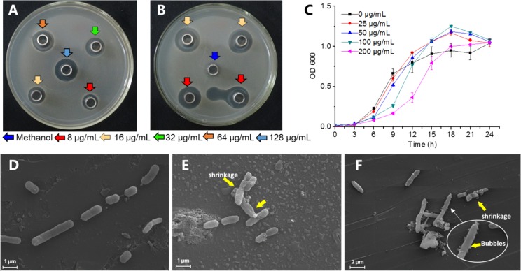
Figure 5.
Biomarker γ-mangostin inhibited the growth of R. solanacearum. (A) Antibacterial studied using the Oxford cup method on the agar medium at different concentrations of γ-mangostin. (B) Different concentrations of streptomycin sulfate served as positive control against R. solanacearum; methanol used as a negative control. (C) Effect of γ-mangostin on the growth of R. solanacearum in a liquid medium. (D–F) SEM image of R. solanacearum cells treated with γ-mangostin. Panel (D), untreated control; panels (E) and (F), treated with γ-mangostin at final concentrations of 16 and 128 μg/mL, respectively. Yellow arrows show the changes in the cells.

