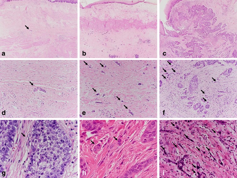
Fig. 1.
Scanning magnification of an ESCC with a subtotal response (regression grade 1B; <10% vital tumour cells) to neoadjuvant therapy (a) showing a good differentiation (CDG-G1) according to the Cellular Dissociation Grade with only a singular cell nest (arrow) within the tumour bed composed of >5 tumour cells (no tumour budding, intermediate cell nest size), which is also shown in higher magnification (d; ×20). Scanning magnification of an ESCC with a marked response (regression grade 2; >10%, <50% vital tumour cells) to neoadjuvant therapy (b) showing a poor differentiation according to the Cellular Dissociation Grade (CDG-G3), with numerous small tumour cell complexes <5 cells (high tumour budding activity; arrows) within the tumour bed and multiple foci of single-cell invasion (smallest cell nest size), which are shown also in a higher magnification (e; ×20). Scanning magnification of an ESCC with a poor response (regression grade 3; >50% vital tumour cells) to neoadjuvant therapy (c) showing a poor differentiation according to the Cellular Dissociation Grade (CDG-G3), with numerous small tumour cell complexes <5 cells (high tumour budding activity; arrows) within the tumour bed and multiple foci of single-cell invasion (smallest cell nest size), which are shown also in a higher magnification (f). High magnification (×40) of tumour budding and cell nest size in 1 HPF: well-differentiated ESCC (CDG-G1; g) without tumour budding and with large cell nest size (>15 tumour cells; arrow). Moderately differentiated ESCC (CDG-G2; h) with low tumour budding (2–4 tumour buds per 1 HPF) and with small cell nest size (2–4 tumour cells; arrows) but without single-cell invasion. Poorly differentiated ESCC (CDG-G3; i) with high tumour budding (≥5 tumour buds per 1 HPF; arrows) and single-cell invasion (arrows)

