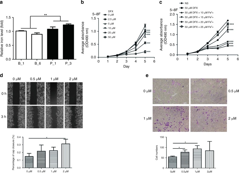
Fig. 6.
Iron retention accelerates cell proliferation, migration and invasion of NPC cells. a Relative concentration of intracellular iron calculated as the total amount of intracellular iron per microgram protein in two sub-colonies each of BDH2–5–8F and pCMV6-Entry–5–8F cells. b MTT assay of proliferation of 5–8 F cells under deferasirox (DFX) treatment. c Proliferation of cells after adding iron (II) sulfate heptahydrate to saturate the iron-chelating ability of deferasirox (50 μM). Data are mean ± SD (n = 5). d Wound-healing assay was performed in 5–8 F cells treated with iron (II) sulfate heptahydrate at 0, 0.5, 1 and 2 μM, respectively. The wound width was measured at 0 and 3 h. Magnification × 100. e Invasive capacity of 5–8F cells, with iron supplement, was assessed by Transwell assay. The number of invading cells was counted. Data are mean ± SD (n = 3). *P < 0.05; **P < 0.01; ***P < 0.001

