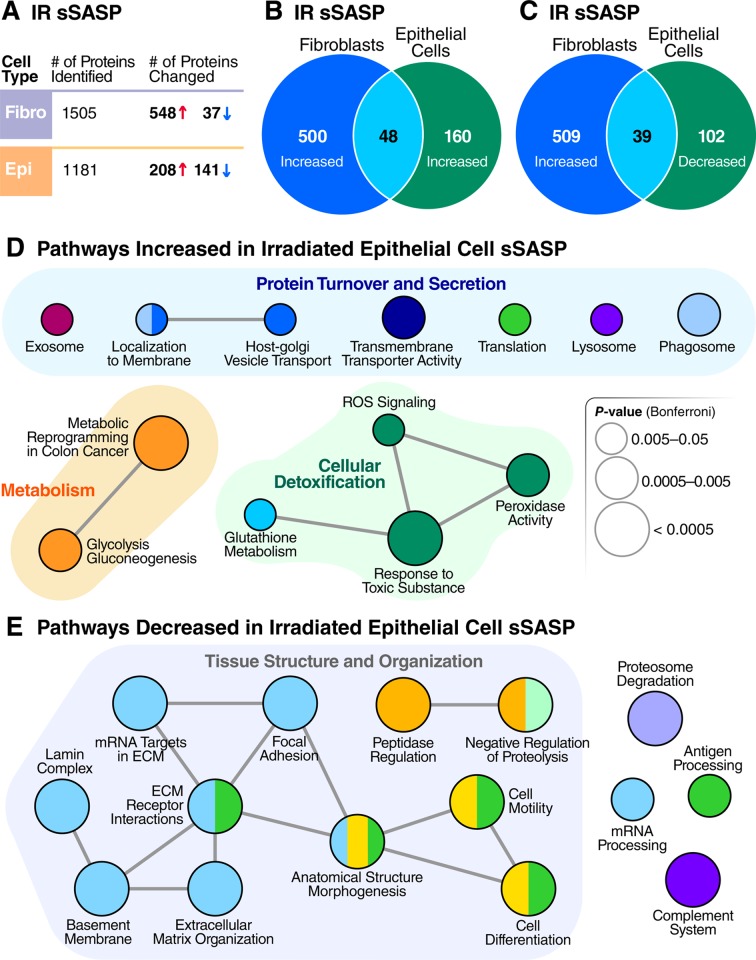
Fig 4. Epithelial cells and fibroblasts exhibit distinct sSASPs.
(A) Number of proteins identified and significantly altered in the sSASP of irradiated fibroblasts and epithelial cells. (B) Venn diagram comparing proteins significantly increased in the sSASPs of senescent fibroblasts and epithelial cells, both induced by IR (q < 0.05). (C) Venn diagram comparing protein increases in the fibroblast sSASP versus decreases in the epithelial sSASP. (D) Pathway and network analysis of secreted proteins significantly increased in epithelial cell sSASP. (E) Pathway and network analysis of proteins significantly decreased in the epithelial cell sSASP. ECM, extracellular matrix; IR, X-irradiation; ROS, reactive oxygen species; sSASP, soluble senescence-associated secretory phenotype.

