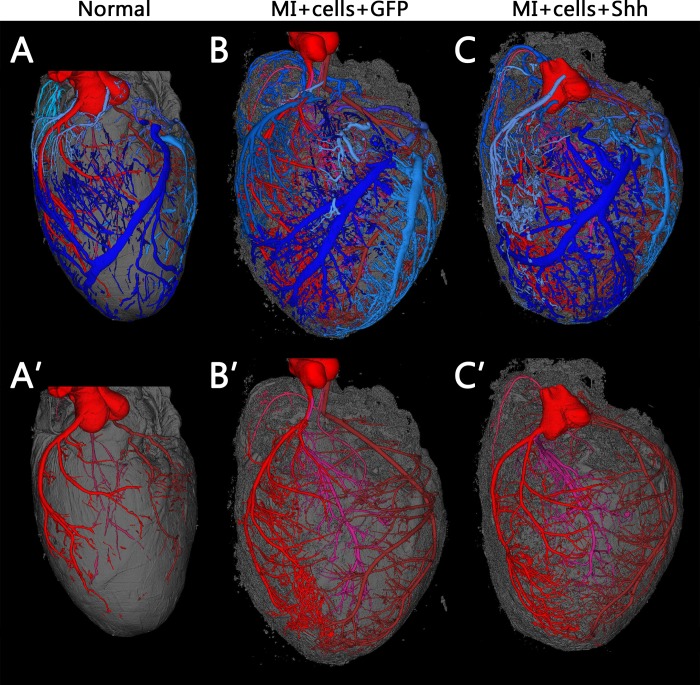
Fig 6. Coronary vascular trees are not visibly different between Shh and GFP treated hearts post-cell engraftment.
Typical 3D reconstructions of coronary vasculature in normal uninfarcted hearts (A) versus hearts that were infarcted and injected with hSC-CMS after treatment with either GFP virus (B) or Shh virus (C). Vessels are segmented and false-colored such that arterial networks are shades of red, venous are blue. Myocardial tissue is shown in gray. The top panels show all vessels and are also available as S1–S3 Videos. Bottom panels show only arterial networks. Both groups of infarcted hearts show hypertrophy and an increase in vasculature visible by μCT, but there is no obvious visible difference between hearts treated with GFP or Shh.

