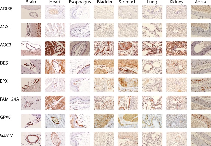
Fig 1. Smooth muscle protein expression in mouse tissues.
All antibodies against listed proteins were first validated on human brain as localizing to cerebral vascular smooth muscle. The same antibodies were used on mouse tissues shown here. With few exceptions, protein was localized to mouse cerebral vascular smooth muscle (first column on the left) and to both vascular and non-vascular smooth muscle in other tissues. In lung, non-vascular smooth muscle around the airways was analyzed. In bladder and stomach, non-vascular smooth muscle is shown in the same field as arteries to serve as a comparison for staining intensity on both cell types. The brain, heart, and aorta were viewed at 400X and all other tissues were viewed at 200X; the scale bar is 100 microns for all tissues.

