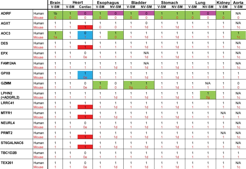
Fig 3. Summary of IHC staining of human and mouse tissues.
We scored for the presence of significant and consistent staining using online data from the Human Protein Atlas for indicated human tissues. We scored mouse tissues stained for the same molecules in a blinded fashion. The presence of protein staining is indicated by 1, while the absence is indicated by 0. N/A indicates that staining was equivocal or inconsistent between samples or that the data was not available. Color codes and letters represent cases of non-conservation between cell types or species that are as follows: purple highlights show proteins expressed in VSMC but not in non-vascular SMC of the same organ in humans only; green highlights show proteins with clearcut differences in cellular staining between human and mouse tissues; blue highlights show proteins that exhibit differences in cardiomyoctes between human and mouse tissues; and red highlights show proteins that exhibited heterogeneity in cardiomyocytes. a: decreased in brain (vs. peripheral) VSMC, b: increased in brain (vs. peripheral) VSMC, c: heterogeneous in VSMC, d: heterogeneous in non-vascular SMC, e: weak, equivocal. V-SM indicates VSMC; NV-SM indicates non-vascular SMC.

