
Figure 1.
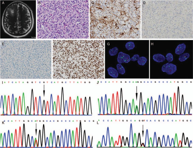
Representative clinico-neuropathological images of patients with lower-grade gliomas. (A) A patient (No. 9) with IDH2 mutant glioma showed a high-signal space-occupying lesion was on right frontal lobe on axial T2-weighted image of MRI. (B) The oligodendroglioma features of “fried eggs” nucleus and “chicken claw” vessels were observed, and mitotic figures were common (hematoxylin and eosin staining, original magnification ×400). (C–F) Immunohistochemical staining, original magnification ×400; tumor cells were immunopositive for GFAP (C), negative for IDH1 R132H (D) and p53 (E), and immunopositive for ATRX (F). (G and H) The tumor cells were harboring 1p/19q co-deletion detected by FISH (G: 1p loss of heterozygosity, H: 19q loss of heterozygosity; original magnification ×1000). (I and J) Sanger sequencing showed tumors were IDH1 wild-type (I: arrow, IDH1 wild-type) and IDH2 mutation (J: arrow, IDH2 R172K, AGG>AAG). (K) A patient (No. 11) showed IDH2 R172W mutation (Sanger sequencing, arrow, IDH2 R172W, AGG>TGG). (L) A patient (No. 12) showed IDH2 R172S mutation (Sanger sequencing, arrow, IDH2 R172S, AGG>AGT). ATRX: Alpha-thalassemia X-linked mental retardation; FISH: Fluorescence in situ hybridization; GFAP: Glial fibrillary acidic protein; IDH: Isocitrate dehydrogenase.