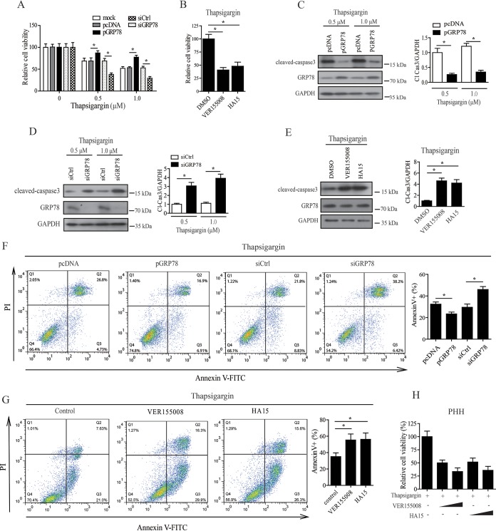
FIG 7.
GRP78 acted as a prosurvival factor in HBV-replicating cells. (A) GRP78 overexpression or knockdown cells were treated with 0.5 or 1 μM thapsigargin for 24 h, and the cell viability was then determined by CCK8 assay. The data are means ± the SEM of five samples pooled from three independent experiments. *, P < 0.05. (B) HepAD38/Tet-off cells were treated with VER155008 (20 μM) or HA15 (10 μM) in the presence of 0.5 μM thapsigargin for 24 h, and the cell viability was determined as in panel A. The data are means ± the SEM of four samples pooled from three independent experiments. *, P < 0.05. (C) HepAD38/Tet-off cells with GRP78 overexpression were treated with 0.5 or 1 μM thapsigargin for 24 h. The cells were then subjected to Western blotting with antibodies against cleaved caspase-3 (Cl. Casp-3), GRP78, or GAPDH. (Right panel) The relative level of Cl. Casp-3 to GAPDH was examined by densitometric analysis, and the value from empty vector-transfected cells was set at 1.0. The data are means ± the SEM of five samples pooled from three independent experiments. *, P < 0.05. (D) GRP78 knockdown HepAD38/Tet-off cells were treated with 0.5 or 1 μM thapsigargin for 24 h, cells were then subjected to Western blotting as in panel C. The data are means ± the SEM of four samples pooled from three independent experiments. *, P < 0.05. (E) Cells were treated as in panel B and then subjected to Western blotting as in panel C. The data are means ± the SEM of three samples pooled from three independent experiments. *, P < 0.05. (F) GRP78 overexpression or knockdown cells were treated with 0.5 μM thapsigargin. The cells were then subjected to apoptosis analysis by fluorescence-activated cell sorting (FACS). The data are means ± the SEM of five samples pooled from three independent experiments. *, P < 0.05. (G) Cells were treated as in panel B and then subjected to apoptosis analysis by FACS. The data are means ± the SEM of four samples pooled from three independent experiments. *, P < 0.05. (H) PHHs were infected with HBV as in Fig. 1D and E. The cells were treated with increasing doses of VER155008 (10 or 20 μM) or HA15 (5 or 10 μM) in the presence of 0.5 μM thapsigargin. The cell viability was determined by a CCK8 test. The data are means ± the SEM of three samples pooled from three independent experiments. *, P < 0.05.

