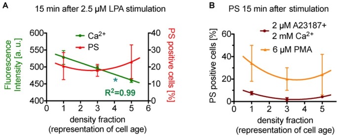
Figure 2.
Reanalysis of data initially presented in Wesseling et al. (2016b). In the original publication, all fractions were only compared in pairs, while here we followed the approach to plot (and analyze) the measured effect in dependence of the cell age. (A) Presents the situation after 15 min of stimulation with lysophosphatidic acid (LPA). While the Ca2+ concentration seems to relate inversely proportional to erythrocyte age (slope is significant different from zero, p < 0.05 is marked with *), phosphatidylserine positive cells show a rather quadratic dependence on cell age. (B) Presents exclusively the phosphatidylserine exposure under more direct stimulations (15 min), namely the direct increase in intracellular Ca2+ in all cells by application of the Ca2+ ionophore A23187 in 2 mM Ca2+ containing solutions (dark red circles) and by a direct Ca2+-independent activation of protein kinase Cα (PKCα, orange triangles) by phorbol-12 myristate-13 acetate (PMA, an unspecific activator of conventional and novel PKCs but PKCα is the sole PKC of these 2 groups found in erythrocytes). Although different in amplitude, both stimulations result in a quadratic dependence on cell age. Blood samples were given from healthy sportsmen (Department of Sports Medicine) with an age between 18 and 36 years. Each data point consists of three donors and for each donor and condition 90,000 cells were analyzed by flow cytometry. For a complete dataset on the different stimulation modes, see Wesseling et al. (2016b). (A) A reprint from Bernhardt et al. (2019).

