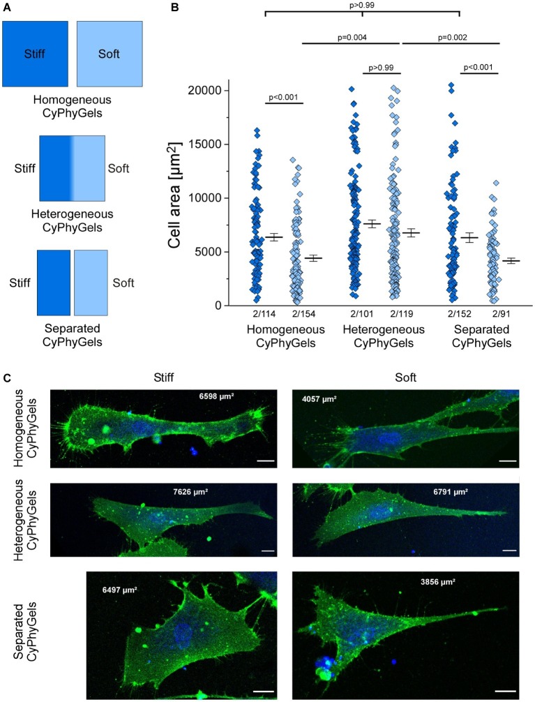
Figure 3.
Area of human primary atrial fibroblasts after 4 days of culture on CyPhyGels in different configurations. (A) Schematic presentation of CyPhyGels used for the experiments in Figures 3, 4. (B) Fibroblasts grown on homogeneously soft CyPhyGels spread less than those grown on homogeneously stiff CyPhyGels. The same applies to separated CyPhyGels, while no difference was found among fibroblasts grown on heterogeneous CyPhyGels (Number of patients/number of cells). (C) Representative images of fibroblasts on different CyPhyGels. Green = cell membrane. Blue = nuclear counterstain. Scale bars = 20 μm (Number of patients/number of cells).

