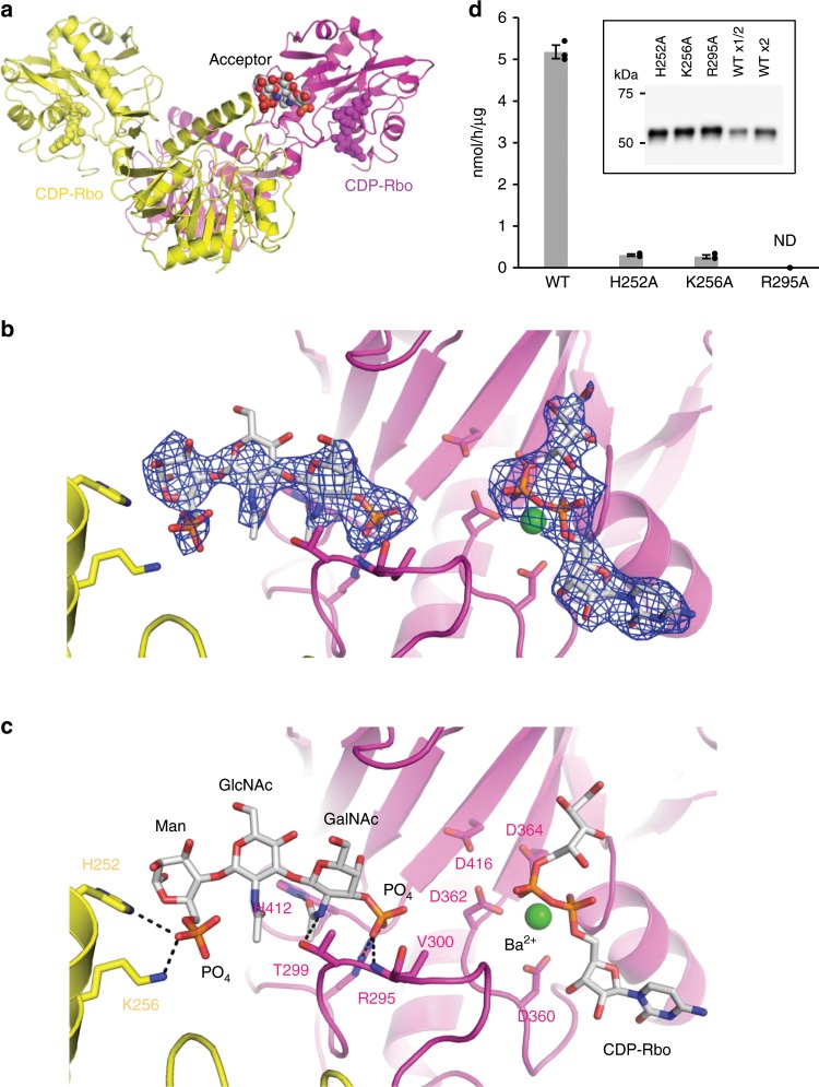
Fig. 4. Structure of sFKRP in complex with the acceptor glycopeptide.
a Two sFKRP subunits are shown in yellow and purple, respectively. CDP-Rbo (yellow and purple) and the phospho-(phospho-)core M3 moiety (CPK color) are shown by sphere models. b Fo-Fc omit map around phospho-(phospho-)core M3 (left) and CDP-Rbo (right). The contour level of the map is 3.0 σ. c Interaction between the protomeric dimer and phospho-(phospho-)core M3 moiety. The trisaccharide is shown as a stick model and interactions between sFKRP are depicted as dotted black lines. d Enzymatic activities of sFKRP (WT, H252A, K256A, and R295A) with CDP-Rbo and the RboP-(phospho-)core M3 peptide. ND, not detected. Average values ± SE of three independent experiments are shown. Each dot represents one data point. Inset: immunoblot analysis of sFKRP proteins immunoprecipitated from the culture supernatant to normalize input sFKRP. Source data are provided as a Source Data file.

