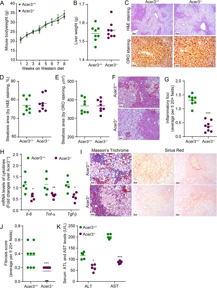
Fig. 2. Acer3 deficiency attenuates early inflammation and fibrosis in the liver of mice with NASH.
a and b Acer3+/+ and Acer3−/− mice were fed PEWD for 8 weeks and their body weights were recorded weekly a. At week 8, mice were euthanized and their liver weights were measured b. The liver tissues were subjected to the following assays. c–e Liver tissues were sectioned for H&E and ORO staining c. Liver sections stained with H&E d or ORO e were analyzed for steatosis areas as described in “Materials and methods” section. f and g Liver sections were stained with H&E f and hepatic inflammatory foci that marked by red cycle were counted to evaluate inflammatory infiltration g. h Total RNA was extracted from liver tissues and subjected to qPCR analyses for the mRNA levels of Il-6, Tnf-α, Tgf-β, and β-Actin as a reference gene. i and j Liver sections were stained with Mason’s trichrome and Sirius Red i and fibrosis was scored as described in “Materials and methods” section j. k Whole blood was collected from Acer3+/+ and Acer3−/− mice fed PEWD for 8 weeks and serum levels of ALT and AST were determined to evaluate hepatocellular injury. Data in a represent mean ± SD, n = 8. Images in c, f, and i represent results from one of eight pairs of mice. *P < 0.05, **P < 0.01, ***P < 0.001.

