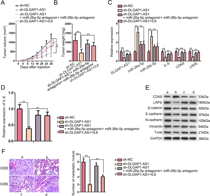
Fig. 8. DLGAP1-AS1 contributed to HCC growth and metastasis in vivo.
a Tumor volume from different treatment groups was measured after injection. b Tumor weight from different treatment groups was measured after the mice were euthanized. c–e The expression levels of several genes related with the present study from each group of xenograft were evaluated using qRT-PCR, ELISA and WB. f Representative images of HE-stained mouse lung tissues were taken to demonstrate lung metastasis of HCC xenograft. The amount of metastatic nodules from each group was calculated and analyzed accordingly. All data are presented as the mean ± SD of three independent experiments. *p < 0.05, **p < 0.01.

