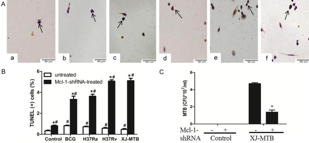
Figure 2.
Mcl-1-shRNA treatment increased apoptosis of mouse peritoneal macrophages infected with BCG, H37Ra, H37Rv and XJ-MTB (×400) and reduced the intracellular growth of MTB. A and B: Apoptosis of Mcl-1-shRNA-treated mouse peritoneal macrophages infected with different strains. TUNEL assays of mouse peritoneal macrophages Apoptotic cells were observed to be dark brown under light microscopy, while normal cells were not stained. a: Control group; b: Mcl-1-shRNA treatment group; c: Mcl-1-shRNA+BCG; d: Mcl-1-shRNA+H37Ra; e: Mcl-1-shRNA+H37Rv; f: Mcl-1-shRNA+XJ-MTB. Percentages of cells undergoing apoptosis. Data represent the mean ± SD of three independent assays in each experiment. *P<0.05 for Mcl-1-shRNA-treated group compared with untreated group. #P<0.05 for infected group compared with uninfected group. Values are the mean ± SD. C: CFUs were recovered from 107 untreated host cells or from Mcl-1-shRNA-treated host cells that were infected with XJ-MTB. Data represent the mean ± SD of three independent assays plated in triplicate. *P<0.05 for XJ-MTB-infected host cells compared with Mcl-1-shRNA-treated XJ-MTB-infected cells.

