
Figure 1.
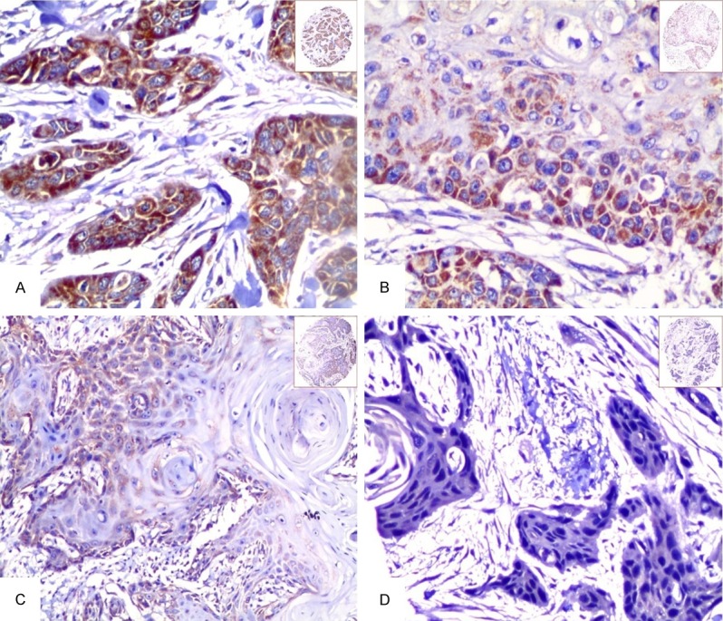
Expression of Hsp10 in OSCC tissue was detected by IHC. The expression of Hsp10 was detected by IHC using specific antibodies as described in the section of materials and methods. Strong positive staining of Hsp10 was identified in the cytoplasm of OSCC (A, 20×, IHC, DAB staining). Moderate positive staining of Hsp10 was showed in the cytoplasm of OSCC cells (B, 20×, IHC, DAB staining). Weak positive staining of Hsp10 was localized in the cytoplasm of OSCC cells (C, 20×, IHC, DAB staining). Negative staining of Hsp10 was in the OSCC cells (D, 20×, IHC, DAB staining).