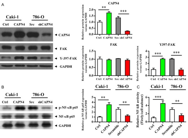
Figure 2.
Capn4 activates FAK and NF-κB signaling pathways in RCC cells. A. Western blot analysis revealed that the phosphorylation level of FAK is up-regulated in the Caki1-Capn4 group or down-regulated in the 780-O-shCapn4 group compared to the respective control cells. B. Western blot analysis revealed that the phosphorylation level of NF-κB is up-regulated in the Caki1-Capn4 group or down-regulated in the 780-O-shCapn4 group compared to the respective control cells. C. Caki1 and 780-O cells were transfected with an NF-κB-dependent reporter plasmid (pBVIx-Luc) and NF-κB activation was determined by measuring relative luciferase activity 48 hours after treatment. Luciferase activity is reported as arbitrary relative light units. Data are shown are mean ± SEM from three independent experiments. Statistical significance was assessed by Student’s t-test; **P<0.01, ***P<0.001.

