
Figure 2.
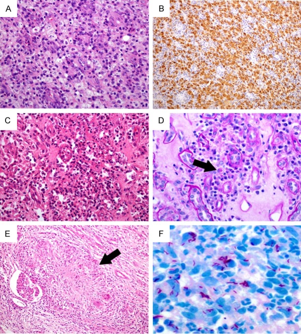
The histopathological changes of case 1, 3 and 4. In case 1, the inflammatory infiltration of xanthogranulomatous pyelonephritis showing variable numbers of foamy histiocytes (A: Hematoxylin & eosin, 200×) with strong and diffuse CD68 immunostaining (B: 100×). In case 3, renal malakoplakia exhibiting aggregates of macrophages, focal microabscesses (C: Hematoxylin & eosin, 200×), Michaelis-Gutmann body within the cytoplasm of the histiocyte (D: Black arrow, Periodic Acid Schiff staining, 400×). In case 4, renal tuberculosis presenting as epithelioid granuloma (E: Black arrow, hematoxylin & eosin, 100×) and Ziehl-Neelsen staining revealing acid fast bacilli (F: 600×).