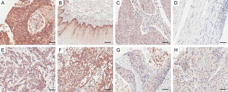
Figure 1.

Immunohistochemical expression patterns of Occludin. A. The strong staining intensity of Occludin. B. The moderate staining intensity of Occludin. C. The weak staining intensity of Occludin. D. The negative staining intensity of Occludin. E, F. The high expression of Occludin in ESCC. G, H. The low expression of Occludin in ESCC. The scale bar represented 50 µm.
