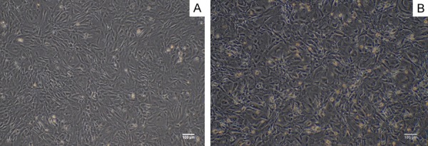
Figure 1.

Characterization of MEF cells. A. The primary MEF cells showed a long spindle shaped, cytoplasmic full and strong in stereo dimension. Bar = 100 micrometer. B. Mitomycin C-inactivated MEF as a feeder layer cells: the cell became slender and some black particles uniformly distributed in the cells. Bar = 100 micrometer.
