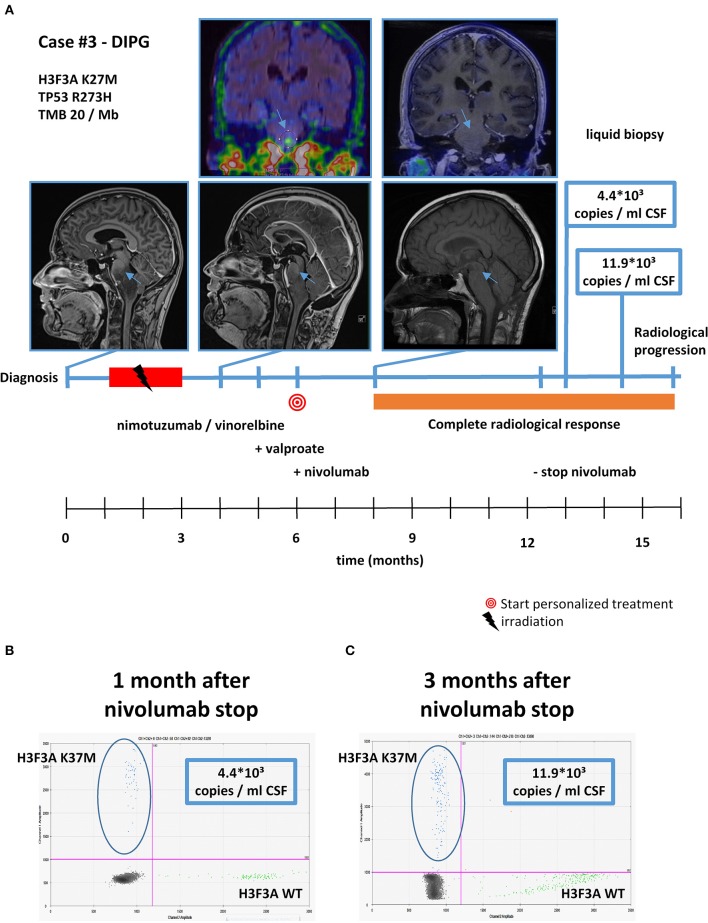
Figure 6.
Detailed case description—case #3. (A) Timeline of patient treatment including magnetic resonance images, 18F-FET-PET images, and liquid biopsy results at indicated time points. Blue arrows indicate tumor. (B) Digital droplet PCR (ddPCR) plot in cerebrospinal fluid (CSF) 1 month after discontinuation of nivolumab. (C) ddPCR plate in CSF 3 months after discontinuation of nivolumab. DIPG, diffuse intrinsic pontine glioma.

