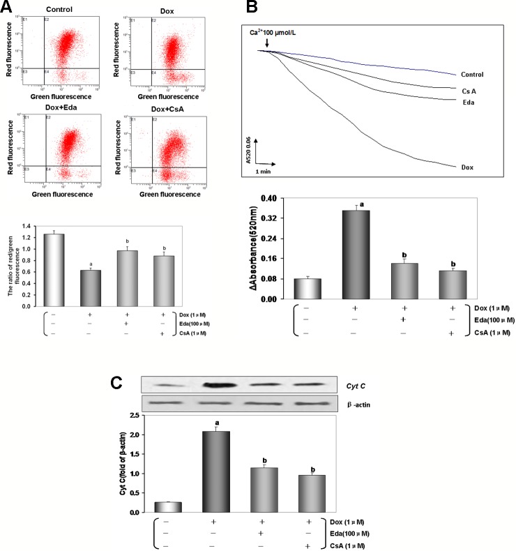
Figure 8.
Dox toxicity could induce mitochondrial dysfunction in human umbilical vein endothelial cells (HUVECs). (A) Mitochondrial membrane potential (MMP) level was evaluated by JC-1. The ratio of red to green fluorescence intensity of cells reflected the level of MMP. (B) Ca2+-induced swelling of mitochondria was used to determine mPTP opening. The changes in absorbance at 520 nm were detected every 2 min. The data were accessed by the following equation: ΔOD = A5200min-A52020min. (C) Western blot analysis and histogram of cyt c expression in cytosol. From left to right, lane 1: control; lane 2: Dox; lane 3: Eda+Dox; lane 4: CsA+Dox. Data are presented as the mean ± S.E.M. for eight individual experiments. a: P < 0.01, vs. the control group; b: P < 0.01, vs. the Dox group.

