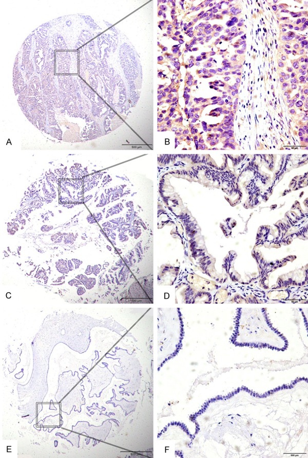
Figure 2.

Expression of LAMP1 in EOC with tissue microarray (TMA). Positive staining was predominantly localized in the cytoplasm of EOC cells. LAMP1 protein expression in tumors from EOC patients showed three different levels. (A and B) Showed strong IHC staining of LAMP1 in EOC samples with advanced cancer, expressing high LAMP1 levels. (C and D) Showed weak IHC staining in EOC samples, (E and F) Showed negative IHC staining of LAMP1. Original magnification ×40 in (A, C, E) (scale bars 500 μm); ×400 in (B, D, F) (scale bars 50 μm).
