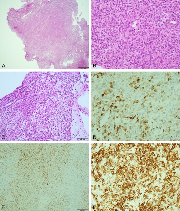
Figure 1.

Specimen obtained by biopsy. (A) Hematoxylin and eosin (HE) (40 ×) (bar is 500 μm), (B) HE (400 ×) (bar is 50 μm), (C) HE (200 ×) (bar is 100 μm). The tumor was immunopositive for keratin (D), S-100 protein (E), α-smooth muscle actin (F) (400 ×). (D-F) Bar is 50 μm.
