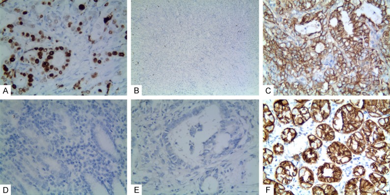
Figure 1.

Positive staining of ORAOV1, or CD133, or WWOX in gastric adenocarcinoma or the control tissue. A: Positive staining of ORAOV1 in nuclei of GAC tissue (100 magnification); B: Negative staining of ORAOV1 in the control tissue (40 magnification); C: Positive staining of CD133 in the membrane and cytoplasm of cancer cells (400 magnification); D: Negative staining of CD133 in the control tissue (100 magnification); E: Negative staining of WWOX in the cancer cells (100 magnification); F: Positive staining of WWOX in the cytoplasm of the control tissue (400 magnification).
