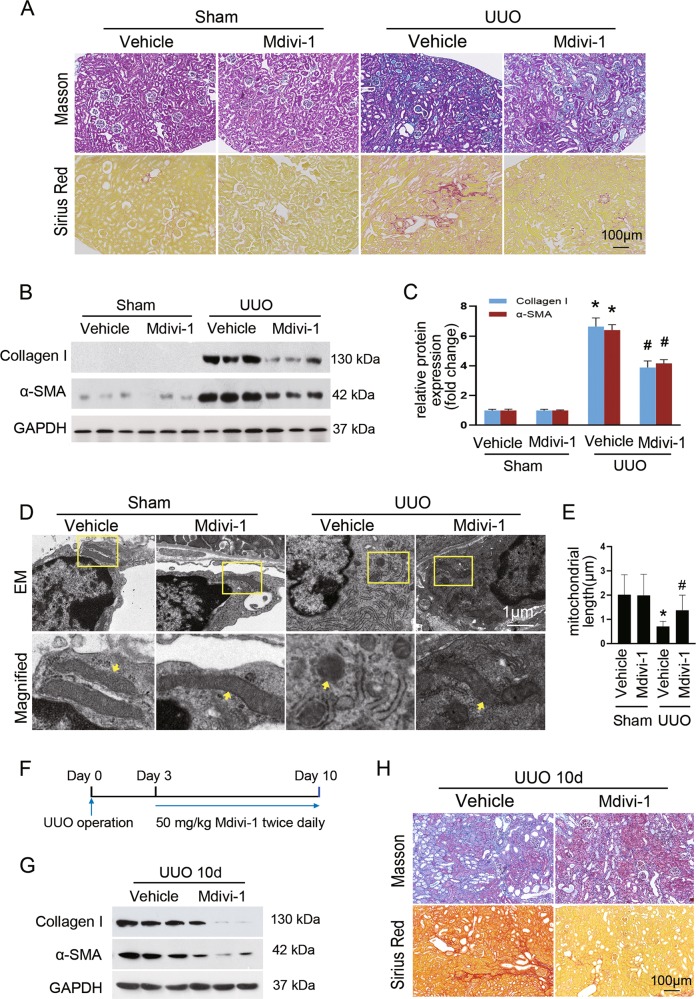
Fig. 2. Suppression of mitochondrial fission by Mdivi-1 alleviates renal fibrosis.
a Masson’s trichrome (upper panels) or Sirius red staining (lower panels) of renal sections among indicated groups. b Immunoblot analyses showing the expression of collagen I and α-SMA in the fibrotic kidneys compared with sham control. c Densitometry analyses of collagen I and α-SMA in immunoblots. GAPDH was used as a loading control. d Representative electron micrographs of mitochondria from renal interstitial fibroblasts among groups as indicated. e Quantitative analysis of mitochondrial length. f Experimental design of evaluating the therapeutic effect of mdivi-1 on established fibrosis. g Immunoblot analyses showing the expression of collagen I and α-SMA in the mdivi-1-treated fibrotic kidneys compared with vehicle control. h Masson’s trichrome (upper panels) or Sirius red staining (lower panels) of kidney sections of the indicated groups. Data in c and e are means ± SEM (n = 6 per group); *p < 0.05 vs sham groups; #p < 0.05 vs vehicle-treated obstructed groups.

