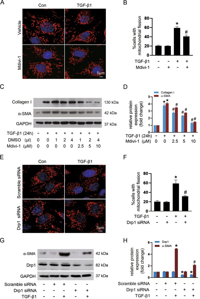
Fig. 3. Targeting Drp1-mediated mitochondrial fission blocks TGF-β-induced fibroblast activation in vitro.
NRK-49F cells were pretreated with Mdivi-1 or transiently transfected with either Drp1 siRNA or scramble siRNA, followed by stimulation with 10 ng/ml TGF-β1 for 24 h. a Representative images of mitochondria stained with MitoTracker Red in cells among indicated groups. b Quantification of the percentage of cells displaying fragmented mitochondria. c Immunoblot analyses of collagen I and α-SMA in cells treated with or without mdivi-1 and exposure to TGF-β1. d Densitometric analysis of immunoblots in c. e Representative images of fibroblasts subjected to MitoTracker (red) and DAPI (blue) staining among indicated groups. f Quantitative analyses for the percentage of cells displaying fragmented mitochondria. g Immunoblot analyses of Drp1 and α-SMA protein expression in fibroblasts among different groups. h Relative expression levels of Drp1 and α-SMA normalized to GAPDH by densitometry. Data in b, d, f, and h are expressed as means ± SEM (n = 3); *p < 0.05 vs TGF-β1-untreated cells; #p < 0.05 vs TGF-β1-treated cells alone or with scramble siRNA.

