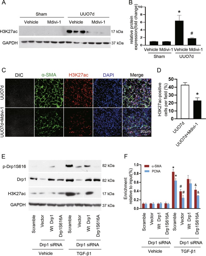
Fig. 6. Drp1 facilitates H3K27ac binding at the promoters of α-SMA and PCNA induced by TGF-β1.
a Kidney tissue lysates were subjected to immunoblot analysis using antibodies against H3K27ac and GAPDH. b The expression level of H3K27ac was quantified by densitometry and normalized with GAPDH. Data are means ± SEM (n = 6 per group); *p < 0.05 vs sham groups; #p < 0.05 vs vehicle-treated obstructed groups. c Representative immunofluorescent staining of α-SMA (green) and H3K27ac (red) in kidneys from mice subjected to UUO operation and treated with or without Mdivi-1. Nuclei (blue) were stained with DAPI. d Quantification of cells with H3K27ac positive. Data are means ± SEM (n = 6 per group); *p < 0.05 vs mice treated with vehicle. e Immunoblot analyses of the indicated proteins from NRK-49F cells after various treatment. f Chromatin immunoprecipitation assay was performed with H3K27ac antibody using nuclear extracts harvested from NRK-49F cells after various treatment. The immunoprecipitated DNA fragments were amplified by PCR using primers specific for α-SMA and PCNA promoter. Data are means ± SEM (n = 3); *p < 0.05 vs vehicle; #p < 0.05 vs TGF-β1-treated cells transfected with scramble siRNA; &p < 0.05 vs TGF-β1-treated cells co-transfected with Wt Drp1 and Drp1 siRNA.

