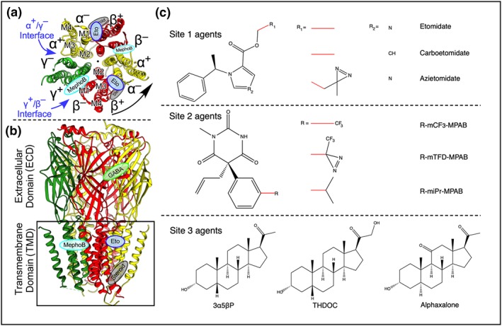
Figure 1.

The structure, binding sites, and agonists of the α1β3γ2 GABAA receptor. (a) Cross section through the TMD viewed from the extracellular side showing previously established drug binding sites and the arrangement of the five subunits around a central pore. (b) Side view; the black box indicates the plasma membrane. Lozenges indicate the binding sites for GABA (green), etomidate (blue), mephobarbital derivatives (cyan), and steroids (grey). The protein backbone is in ribbon notation (Forman & Miller, 2016; Laverty et al., 2019). (c) The chemical structures of agents used in this study
