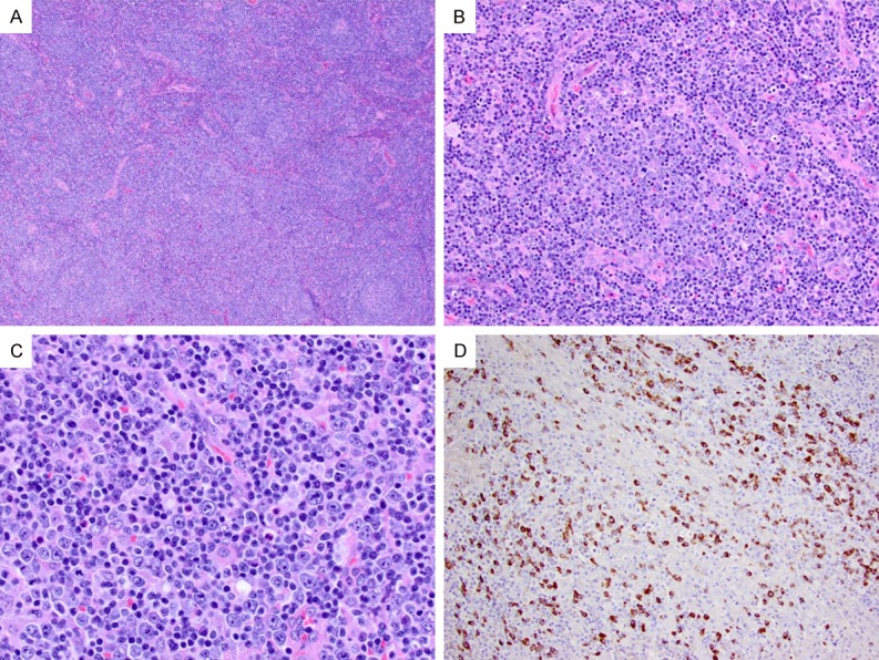
Figure 4.

Type III IgG4-LAD with interfollicular expansion and immunoblastosis. A. The lymph node shows architectural distortion with marked interfollicular expansion (H&E, 40 ×). B, C. The expanded interfollicular regions have frequent immunoblasts, many plasma cells, and scattered eosinophils (H&E, 200 × and 400 ×, respectively). D. Abundant IgG4+ cells are present in the interfollicular areas (200 ×).
