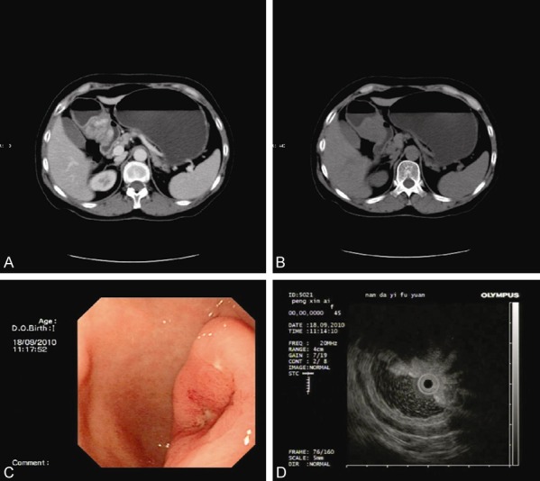
Figure 1.

Whole abdominal CT scan (A and B) shows that there is a shadow of soft tissue mass measuring 20 × 25 mm in size in the antrum. Endoscopy (C) shows that there is an ulcer with a diameter of about 15 mm in the posterior wall of the antrum. Endoscopic ultrasonography (D) findings show that there is a hypoechoic mass in the lesion, with a size of about 22 × 25 mm protruding to the gastric cavity and a less clear boundary. (A) Plain CT scan; (B) Enhanced CT scan; (C) Endoscopy; (D) Endoscopic ultrasonography.
