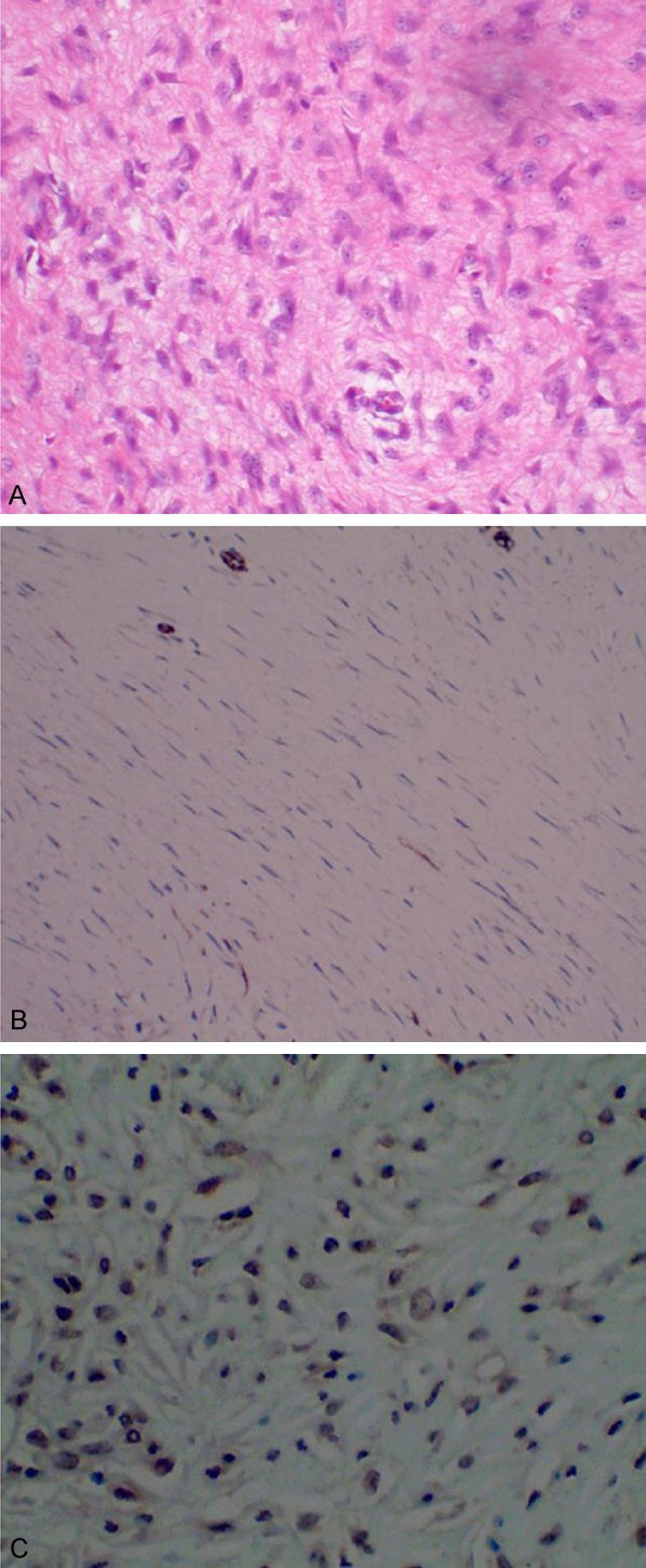
Figure 2.

Microscopic pathological findings (A) indicate the diffuse infiltrative growth of tumor cells in the muscle layer of stomach. Main immunohistochemistry findings include CD34 (-) (B) and β-catenin (+) (C). (A) H&E stain, × 400; (B) CD34, × 400; (C) β-catenin, × 400.
