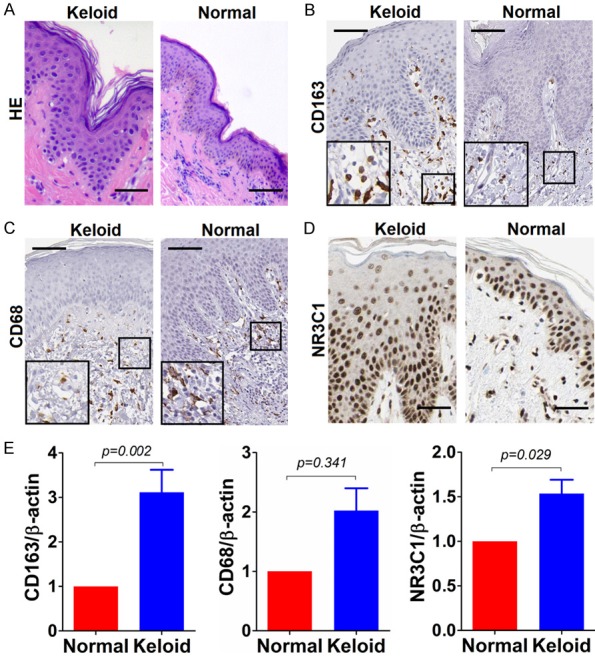
Figure 1.

Immunostaining of CD163, CD68 and NR3C1 in keloid and normal tissues. A. Haematoxylin-eosin (HE) staining of keloid and normal control tissue; B. Immunostaining of CD163 in keloid and normal control tissue; C. Likewise, CD68 staining in keloid and normal control tissue; D. NR3C1 immunostaining in keloid and normal control tissue. Shown were representative figures selected among all cases subjected to immunostaining detection analysis. Scale bar stands for 100 μm. E. Detection of relative expression of CD163, CD68 and NR3C1 on mRNA level using qRT-PCR method in 30 cases of keloids and normal controls. Independent sample T test was used to analyze the difference between keloid and normal control group.
