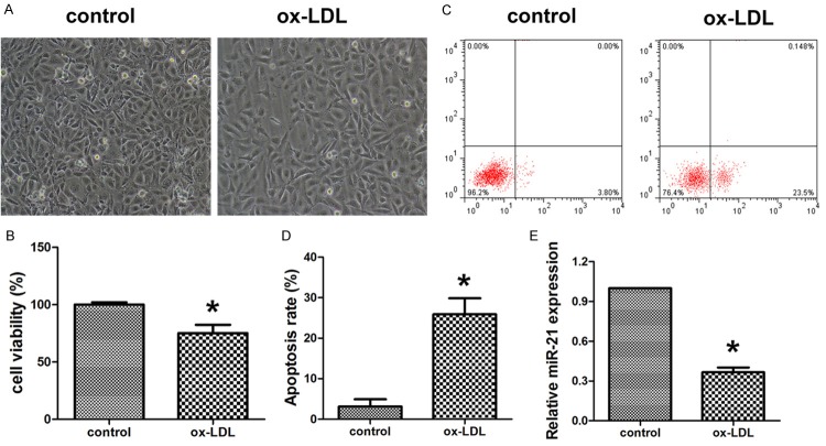
Figure 1.
Ox-LDL induced HUVECs apoptosis and inhibited miR-21 expression. A. The cellular morphology after treatment with ox-LDL and control. B. MTT was used to evaluate the cell viability of HUVECs after treatment with ox-LDL and control. C, D. Flow cytometry was used to detect the apoptosis of HUVECs after treatment with ox-LDL and control. E. Quantitative realtime PCR was applied to evaluate the expression of miR-21. Data are expressed as mean ± SD; n = 8, *P<0.05.

