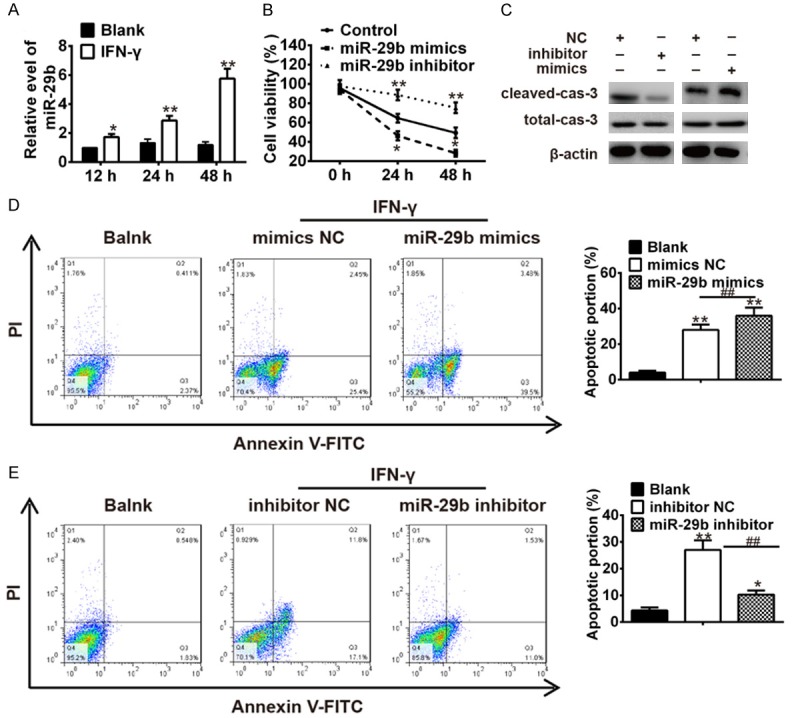
Figure 2.

IFN-γ stimulated keratinocytes apoptosis by up-regulating miR-29b. A. Keratinocytes were treated with IFN-γ (10 ng/ml for 12 h, 24 h and 48 h), and then miR-29b was measured by qRT-PCR. *P<0.05, **P<0.01 vs. Blank group. IFN-γ treated keratinocytes were transfected with miR-29b mimics, miR-29b inhibitor and miRNA negative control. B. Cell viability was measured by MTT assay. *P<0.05, **P<0.01 vs. Control group. C. The expressions of cleaved-caspase-3 and total caspase-3 were analyzed by Western Blot. D and E. The apoptosis was measured by flow cytometer. *P<0.05, **P<0.01 vs. Blank group, ##P<0.01 vs. inhibitor NC or mimic NC. Data are presented as mean ± SD from three independent experiments.
