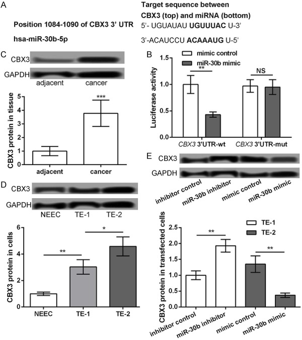
Figure 3.

CBX3 as a target of miR-30b. A: The predicted sequence of CBX3 and miR-30b. B: Luciferase reporter assay showing the luciferase report activity of CBX3 3’UTR-wt and CBX3 3’UTR-mut after transfection with miR-30b mimic or mimic control. C: Western blot showing the protein expression level of CBX3 in ESCC tissues and their adjacent normal tissues. D: Western blot showing the protein expression level of CBX3 in ESCC cell lines (TE-1 and TE-2) and normal esophageal cell line NEEC. E: TE-1 cells transfected with miR-30b inhibitor and inhibitor control, while TE-2 cells transfected with miR-30b mimic and mimic control. The Western blot showing the protein expression level of CBX3 in different transfected groups. *P<0.05, **P<0.01 and ***P<0.001 compared with corresponding control.
