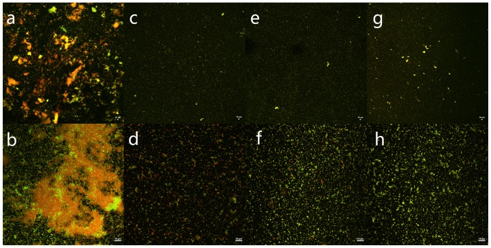
Figure 3.
Confocal laser scanning microscopy (CLSM) images displaying the S. aureus biofilm. SYTO9 and PI interacted with the nucleic acids of live bacteria and dead bacteria, which are shown as green and red, respectively. (a, c, e and g) The biofilm was observed under 10× objective; (b, d, f and h) The biofilm was observed under 60× objective. (a and b) The biofilm was treated with PBS. (c and d) The biofilm was treated with 45 mg/ml immunoliposomes. (e and f) The biofilm was treated with 45 mg/ml liposomes. (g and h) The biofilm was treated with 45 mg/ml ISMN. S. aureus, Staphylococcus aureus; ISMN, isosorbide mononitrate.

