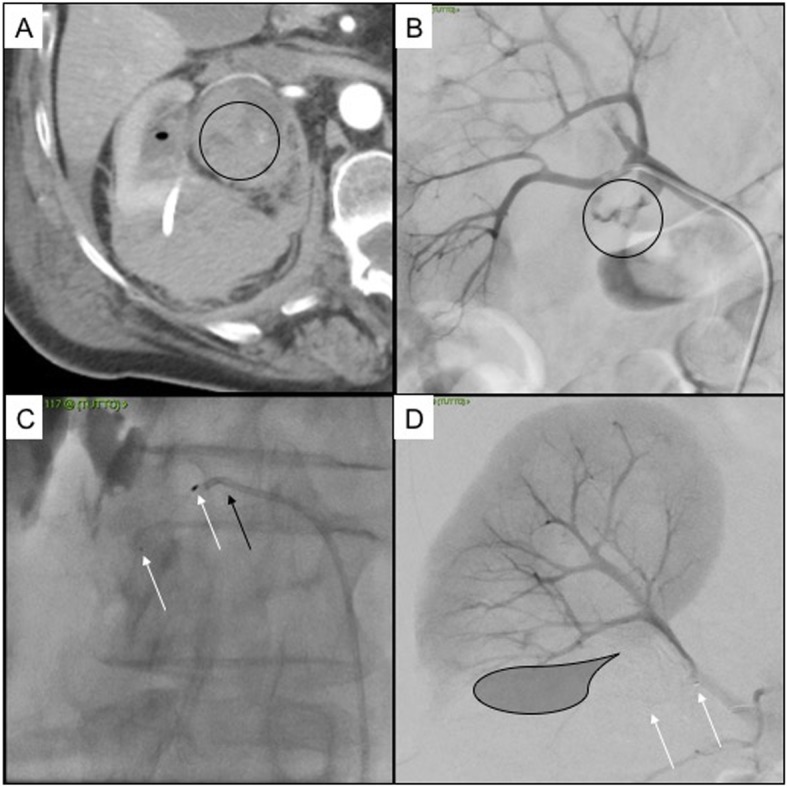
Fig. 2.

Patient 4. 80 years old female with urinary stones causing right pielectasia, treated with nephrostomy. a Axial CT scan in arterial phase showing active bleeding into the urinary pelvis (black circle), clots are appreciable into the calix (black ovoid), the nephrostomy drainage with perirenal hematoma is appreciable as well; b DSA showing extraluminal blush from the inferior segmental artery (black circle); c selective embolization with MVP 7Q (white arrows indicate distal and proximal radiopaque markers of the MVP, black arrow indicates the 5Fr diagnostic Cobra 1 catheter); d final DSA control showing no more angiographic bleeding (black shadow indicates the ischemic area, white arrows indicate distal and proximal radiopaque markers of the MVP)
