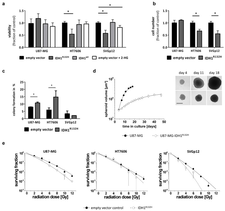
Figure 2.
IDH1R132H reduces growth and increases radio-sensitivity: (a) Cell viability was determined using a WST-1 based colorimetric assay after 48 h culture in adherent condition. Values were normalized to empty vector cells and means from all experiments performed with different transductions were compared (* p < 0.01; one-way analysis of variance (ANOVA) followed by Dunnett’s post-hoc t-test). (b) Quantification of cell numbers counted using CASY® TTC 72 h after seeding. (c) Clonogenic survival assays showed that IDH1R132H significantly enhanced the capacity of glioblastoma cells to form colonies, but not of the astrocytes. (d) To analyze cell growth under 3-D conditions, U87-MG control and IDH-mutant cells were seeded in liquid overlay and cultured for up to 50 days. Spheroid size and volume were routinely monitored. In the example shown here, 2 × 10³ cells/well were seeded. Data are expressed as the mean volume ± SD. Representative phase contrast microscopic images of the same spheroids are shown at day 4, 11, and 18 (scale bare = 400 µm). (e) Radiation-dose response curves were derived from clonogenic survival assays. Data are expressed as mean of three biological experiments ± SD with n ≥4 wells per experiment and treatment condition. Dose response curves were fitted using a linear-quadratic model (surviving fraction = exp − (αD + βD2)).

