Figure 2.
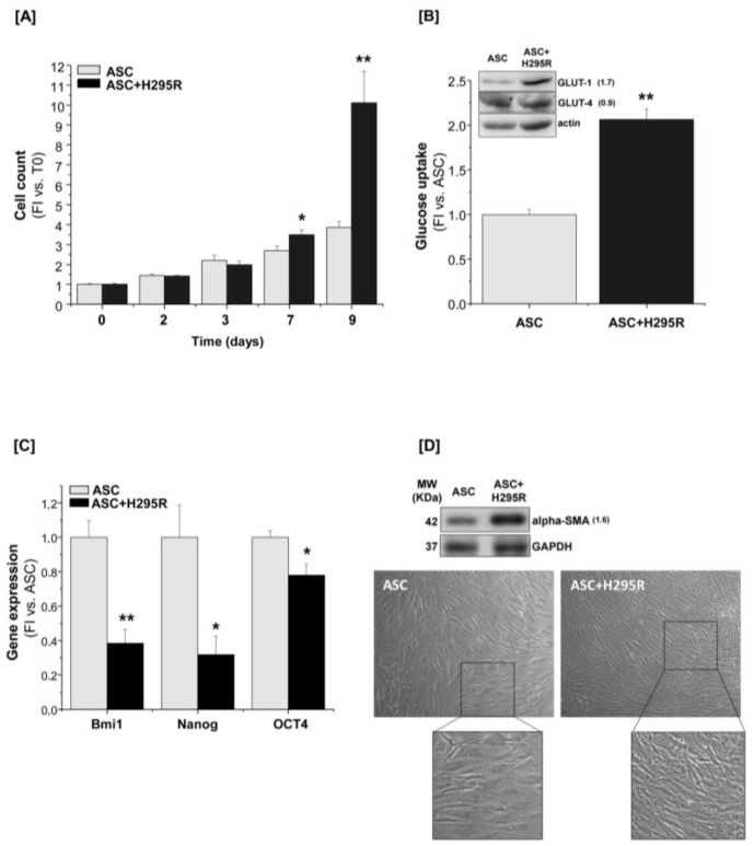
H295R cells stimulate ASC proliferation and drive ASC differentiation toward a myofibroblast-like phenotype. (A) ASCs alone (ASC) or co-cultured with H295R (ASC+H295R) were assessed for cell proliferation at the indicated time points (2, 3, 7 and 9 days) by direct cell count. The proliferative rate was calculated as fold increase (FI) versus the co-culture starting time (Time point = 0), n = 5. (B) Glucose uptake measurement and western blot analysis of GLUT-1 and GLUT-4 expression (inset, fold increase intensity vs. ASC after normalization on actin band is indicated to the right of the bands) assessed in ASCs after 7-day mono- or co-culture, n = 3. (C) Gene expression of specific mesenchymal stem-related markers revealed by RT-qPCR Taqman assay in 7-day co-cultured ASCs compared with the ASC mono-culture, n = 3. (D) Western blot analysis of α-SMA expression and optical microscopy of ASCs cultured alone or in the presence of H295R cells for 7 days. Original magnification: 10×; zoom in: 2×. For western blot analysis, GAPDH or actin were used as internal loading control. Gene expression and glucose uptake are indicated as fold increase (FI) versus ASCs alone. Data are expressed as the mean ± SE in at least three independent experiments; * p < 0.05; ** p < 0.001. Details of western blot can be viewed at the Supplementary Materials.

