Figure 7.
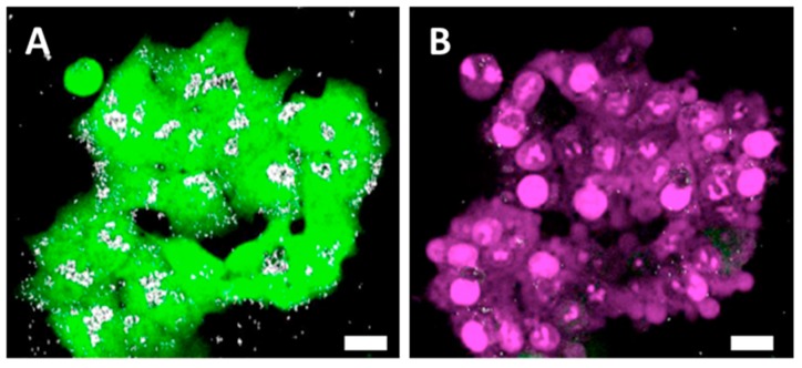
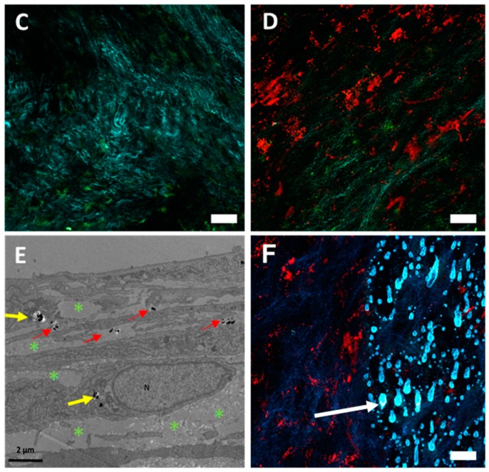
Nanochains as photothermal mediators affecting the cells and the extracellular matrix. (A) HCT-116-GFP cells (green) loaded with nanochains-COOH (white spots), exposed to the laser within the multiphoton microscope. (B) Nanochains-COOH-loaded HCT-116-GFP cells, which underwent cell death and thus internalized propidium iodide after laser exposure (the red fluorescence derived from cell-internalized propidium iodide is here represented by a magenta pseudo-color). (C) Representative micrograph of a control cell sheet exhibiting a rich collagenous matrix (turquoise) among auto-fluorescent (green) fibroblast cells (D) Representative micrograph of a cell sheet loaded with red-fluorescent RB-nanochains-COOH surrounded with collagen (turquoise) and fibroblasts (green). (E) TEM micrograph showing the distribution of RB-nanochains-COOH within the cell sheet. N denotes cell nucleus, yellow arrows point to intracellularly localized nanochains and red arrows point to extracellular nanochains. Green asterisks denote the collagen fibers in the extracellular matrix. (F) Representative micrograph of a cell sheet: exhibiting on the left side the intact collagen fibers (exposed to 20 mW laser power), and on the right side the melted collagen fibers (exposed to 33 mW laser power power). Scale bars in (A–D,F) equal 20 µm. White arrow in (F) points to a characteristic “drop” of melted collagen fibers.


