Figure 1.
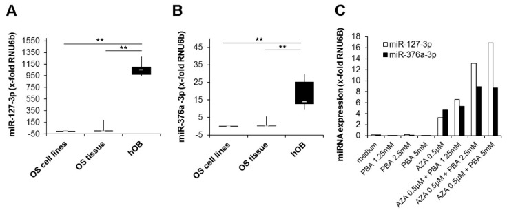
Silencing of miR-127-3p and miR-376a-3p in osteosarcoma cell lines and tissue. Quantitative real-time PCR analysis of (A) miR-127-3p and (B) miR-376a-3p expression in osteosarcoma cell lines (n = 7), osteosarcoma tissue (n = 8), and primary human osteoblasts (hOBs) (n = 5). Expression values were normalized to the expression of the small nuclear RNA U6 pseudogene (RNU6B). The white lines indicate the medians, the lower boundary of the box of the 25th percentile and the upper boundary of the box of the 75th percentile. The whiskers indicate the highest and lowest values. p-values were determined by the Mann–Whitney U test. (** p < 0.01). (C) Upregulation of miRNA expression by epigenetic modifiers. The osteosarcoma cell line 143B was treated for seven days with the indicated concentrations of 5′-Aza-2′-deoxycytidine (AZA) followed by a further three days of culture with or without the addition of phenylbutyric acid (PBA). After the incubation period, the expression of miR-127-3p and miR-376a-3p was quantified and normalized to the expression of RNU6B.

