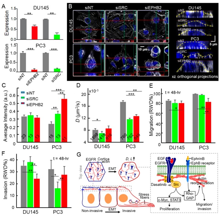
Figure 5.
Disruption of EphB2/Src pathways leads to attenuated cell motility, invasion, and EGFR diffusion in advanced prostate cancer cells. (A) Effective gene knockdowns in siRNA-treated DU145 and PC3. (B) Structured Illumination Microscopy (SIM) images of siRNA treated cells. Maximum intensity projection on the xy plane and orthogonal cross-sections (xz and yz) of DU145 and PC3 siRNA treated cells. (C) Quantification of cortical actin based on fluorescence intensities of xz and yz orthogonal projections along the apical plasma membrane. The number of projections analyzed is labeled on each bar. (D) EGFR diffusivities of the siRNAs treated cells. The error bar represents the standard error of the mean. (E,F) The image-based assays allow us to conduct the time-lapse analysis of cell migration and invasion on the siRNA-treated cells. The error bar represents the standard deviation. All statistical analyses were performed using the unpaired t-test. The asterisk represents the level of statistical significance for t-test: *** p < 0.001, ** p < 0.01, * p < 0.05. (G) Schematic shows the effects of EMT-induced actin reorganization on EGFR dynamics and the Src/EphB2 induced signaling from the plasma membrane that controls cell behavior.

