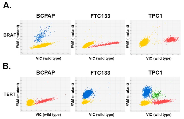
Figure 1.
Detection of BRAF (B-type Raf kinase) and TERT (Telomerase Reverse Transcriptase) mutations by digital PCR in thyroid cancer cell lines. Each panel represents a single dPCR experiment whereby a DNA sample was segregated into individual wells and assessed for the presence of the mutant allele and wild type allele using two different fluorophores (6-Carboxyfluorescein (FAM) and 2′-chloro-7′phenyl-1,4-dichloro-6-carboxy-fluorescein (VIC). The signals from the FAM (blue) and VIC (red) dyes were plotted on the y-axis and x-axis, respectively. The yellow cluster represented unamplified wells (negative calls). (A) 2D plots of dPCR reads out of DNA extracted from thyroid cancer cells. Blue cluster represented wells that were positive for the BRAFV600 mutation. (B) Results of dPCR analysis of TERT228 analysis in thyroid cancer cells, demonstrating the presence of mutant (blue) and wild type (red) alleles. Green cluster represents wells containing both VIC and FAM dyes.

